Advances in technology, like in vitro and microfluidics, have helped advance research
 America’s population is aging, with the number of people over 65 expected to reach 80.8 million by 2040, more than double the number in 2000. This will, unfortunately, also lead to more people developing neurodegenerative diseases, such as Alzheimer’s and Parkinsons. These diseases are already rapidly rising around the world, to the point where over a third of the population is affected by neurological conditions, making it the leading cause of illness and disability.
While it can be frustrating as we wait for a cure, especially as we watch our loved ones, and then sometimes even ourselves, succumb to these diseases, there have been major advancements in technology in the last couple of decades that are getting us much closer to that result.
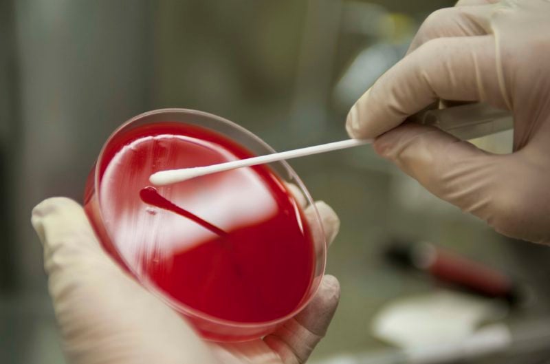
Specifically, in vitro technology, allowing labs to do disease modeling, in which they can create representative systems that mimic the behavior of diseases on human cells rather than on animals. In vitro is Latin for “within the glass”, which makes it an appropriate term for referring to disease modeling outside the body, or in this case, inside a petri dish.
From animal testing to stem cells
The reliance on animal testing has led to a "graveyard of drugs" over the past 30 years as we attempt to cure central nervous system disorders, Thomas Durcan, associate professor at the Montreal Neurological Institute (The Neuro) at McGill University, said to me.
"Drugs come out; they’re the flavor of the month, and then they go into phase two, three trials, and often the data doesn't work the way we would like it to. A lot of it is because the disease is very complex; we're trying to model the disease in animal models and often there's differences between that and what a human is," he said. "That's where the gap has been: how do you take the cells of the brain and really scale experiments and then actually better understand why a disease of the brain is happening or what might be going on with a particular gene, like if it's mutated or affected?"
Thanks to the introduction of in vitro technology, this gap is starting to close. Prior to 2006, research was heavily reliant on animal studies for anything preclinical simply because the barrier to using in vitro, meaning working directly with cell culture, was just too high, and it wasn't necessarily worth it at that point.
It’s been the last five to 10 years, especially, that in vitro has started to become more sophisticated thanks to the number of available cell types that researchers can differentiate stem cells into. The past few years have seen a big focus on figuring out how to make more and more cell types, but also how to use them in a physiologically relevant way. That means, as opposed to studies being done on samples taken from an animal, they can be done on human cells because any cell type of the human body can now be transformed into a stem cell, which can then be made into whatever you want them to be. For example, a cell from the heart or a cell from the liver, giving researchers much more flexibility in regards to what they want to study.
This also makes experiments more predictive: “If drugs are tested in a physiologically-relevant human model, versus inside of an animal, we hope that the results achieved with those experiments will be more useful in terms of how they will work in a patient” said Emma V. Jones, Lead Scientist, In Vitro Neurobiology, at Medicines Discovery Catapult (MDC), an independent, not-for-profit organization and part of the Catapult Network established by Innovate UK. MDC's vision is to reshape drug discovery through partnerships.
"However, it's a really, really difficult challenge, actually, to make something in a laboratory behave and respond and look like something as it would have in a person," she explained.
"Historically, people would have used cell lines, and rodent cells, but in the last 10 years or so there's just been a massive expansion in the use of stem cell derived human models, which is mainly what we're using."
Making cell types accessible
In order to make a more relevant human model, scientists need to have more of the components, including stem cell technologies, which are essential to make the cell types more accessible. Induced pluripotent stem cell (iPSC) technologies are powerful enough to create the cell types, but also to create cell types with a certain disease, which makes research on these diseases much more accessible.
All of this is beneficial for the patient, Durcan explained, because all they have to do is give blood, which is easy, and something they're very willing to do, especially if it means potentially curing their neurodegenerative disease.
"When patients come in, they actually want to see progress made and so they're willing to give materials, and one of those materials happens to be blood. We can take the blood, make it into stem cells, so you can now have a lot of different patients, let's say, with Parkinson's, that you can now make their cells on a dish, their neurons on a dish, and then you can start to understand why they're getting this disease," he explained.
"If we know why they're getting the disease, then you can actually start to see if there's an intervention that maybe could rescue the phenotype we're seeing on the plate." Phenotype refers to observable characteristics of an organism, like levels of hormones.
Microfluidic devices
Another major technological advancement has been the proliferation of microfluidic devices, which contain a small chip, imprinted with very small channels and reaction chambers, that uses forces such as electrokinetic, capillary, and vacuum to mix and separate liquid samples.
These devices have a number of benefits over conventionally-sized systems, such as allowing the analysis and use of lower volumes of samples, chemicals, and reagents, making applications less expensive.
"Microfluidics devices can test disease mechanisms that you can't test using standard systems. These devices open up the opportunity to test disease mechanisms in vitro that previously you couldn't do. It also reduces the use of animals because if we're able to do more complex experiments in vitro, it means that we can reduce the types of experiments that are done in vivo using animals," said MDC's Jones.
These microfluidic devices add an extra layer of complexity when compared to the standard 96-well microplates, where all the cells are sitting together rather than separated. With microfluidics, you can isolate two different cell types, meaning that if you have neurons on one side and another cell type on the other, you can add something to one side and then look at how it spreads to the other side. You wouldn't be able to do that in a 96-well plate because it would all be in the same liquid. "In a 96-well plate cells are all jumbled up together, they're all sitting in the same media, so it's not like you can go into one cell type in your culture and specifically stimulate that or look at the responses produced by that cell," explained Eve Corrie, Scientist, In Vitro Neurobiology, at MDC.
"We're also using microfluidics to model synapses - the place where neurons connect and communicate with each other - in situations where the neuron cell body is really far away from its terminal (end point). In the body, the largest brain cell is like a meter long from the cell body to the end of its axon, or projection of the nerve cell. So how do you model that in a 96-well plate where it's all together?" she said. To give an analogy, axons are like the electrical wires that connect neurons together, and synapses are the transfer points between them.
"In microfluidics, you can separately have your axon and your cell bodies, and you can treat them separately and you can look at the responses separately."
The eNuvio solution
One company in the microfluidic space is eNuvio, which has developed devices that can simulate the body so researchers can bypass animal testing.
These devices are used by both The Neuro and MDC; The Neuro struggled four hours a day changing the media, or the solution that organoids were grown in. Organoids are small pieces of organs grown in a lab. By using eNuvio for the past six years, The Neuro avoided all of that manual labor because the changing of the solution was automated in the eNuvio system. Instead of taking a few weeks to make organoids, eNuvio enabled them to make the same amount within a week and this scale has led The Neuro to many advances, particularly in the area of Parkinson’s.
“Because of these devices, we were able to actually see if we could take these stem cells, make brain organoids, and actually model features of Parkinson's in a dish. Then, gradually, we were able to then do proteomics studies with them," said The Neuro’s Durcan. Proteomics is the study of an organism's entire protein, which is used to diagnose disease and discover drug targets to overcome drug resistance.
“We’ve actually been able to study a lot of the genes implicated in Parkinson's, understand why it's happening, and do it in the context of a model that's almost the closest you'll ever get to what you'll see within a human brain.” While other companies offer microfluidics solutions, eNuvio helped The Neuro reduce hours of daily manual labor.
MDC, meanwhile, uses eNuvio’s devices to model situations in the body where cells are separated from each other, something that would be difficult with co-cultured cells, or other disease mechanisms, such as the spread of pathogenic aggregated proteins.
"We’ve used eNuvio's microfluidics in order to get a degree of spatial separation in our cultures, and fluidic isolation as well, because you can make sure the fluid is only going from one direction to the other," Corrie said. "It sets up a whole new assay with protein seeding in the context of neurodegeneration, and that's completely new for us because of these microfluidic devices." Cell seeding simply means spreading those cells in a vessel (in this case a microfluidic device) so they can be used in culture experiments..
"Before, we couldn't model the spreading of pathogenic proteins which are involved in neurodegeneration properly, but now we're able to do that. We're now able to help drug discovery innovators that are developing therapeutics to target that," said MDC’s Jones.
Whereas most microfluidics chambers have closed channels, eNuvio’s have open chambers that make it easier to seed cells in, MDC’s Corrie explained.
“Seeding these cells through the channel is quite tricky to do correctly and once they’re there you don’t have particularly easy access to them because they’re sitting in a sealed channel. The eNuvio ones just have an open chamber so you can just sit your cells there easily and then you can remove or replace the liquid they’re sitting in very easily too,” she said.
MDC also looked into different suppliers of microfluidic devices but there are a few things that they found appealing about eNuvio, the first being that their devices were ready to go: unlike some other devices, where you have to plasma bond glass coverslips on to them, and they don't come just ready to plate your cells into, the ones from eNuvio do and they're sterile. That means you just take them out of the bag, and you put your cells in them.
Conclusion
By allowing researchers to see how drugs interact with human cells rather than on animals, these technologies like in vitro and microfluidics are not only giving them more control over which cells they’re working with, making experiments more accurate, but they’re also allowing that research to be conducted faster.
These advances are a game-changer for the development of new drugs and therapeutics, and could mean that we could very well finally see a cure to some of these neurodegenerative diseases within our lifetime.
(Image source: pharmaceutical-technology.com)

 Anna Vod